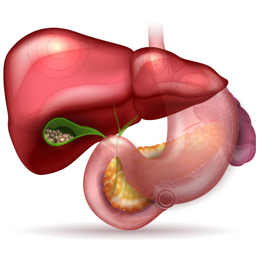
What are gallstones?
Gallstones are stone-like deposits that form in the gallbladder, a small pear-shaped organ located under the liver. The gallbladder is responsible for the storage of bile, a greenish-brown fluid used in the digestive process to digest fats. Bile is made in the liver.
How do gallstones form?
Gallstones develop when crystals form in bile, the liquid stored in the gallbladder. Over time, the crystals can grow into hard, stone-like lumps.
Bile consists of a number of components, including cholesterol, bile salts, bilirubin and water. If your gallbladder fails to empty properly, or if the cholesterol, bile salts or bilirubin become too concentrated, you may develop gallstones.

Gallstones diagnosis
To diagnose gallstones, your doctor will ask about your symptoms, perform a physical examination and recommend some tests. Tests that can detect gallstones and associated conditions include the following.
Ultrasound
Ultrasound scanning is the most common technique used to confirm the presence of gallstones. Ultrasound can:
- detect gallstones in the gallbladder with high accuracy;
- sometimes detect gallstones in the bile ducts (further testing is often needed to confirm stones in the bile ducts); and
- detect signs of gallbladder inflammation.
Ultrasound is a quick and painless procedure that uses high-frequency sound waves, sent through a hand-held device that is moved across the abdomen. The echoes as the sound waves bounce off the gallbladder and other organs are converted to electrical impulses that show a picture on a monitor.
‘Silent’ gallstones, or gallstones that do not cause any symptoms, are sometimes detected incidentally during procedures such as ultrasounds or X-rays.
HIDA scan
A HIDA scan – also known as cholescintigraphy or a hepatobiliary scan – involves having a small amount of a radioactive substance (known as a tracer) injected into a vein and then taking scans.
It can be used to detect:
- obstruction of bile ducts (that can be due to gallstones);
- problems with gallbladder emptying; and
- cholecystitis (inflammation of the gallbladder).
MRI
Magnetic resonance imaging (MRI) produces detailed images of the body, and can be used to:
- visualise the gallbladder; and
- show gallstones in both the gallbladder and the bile ducts.
Endoscopic retrograde cholangiopancreatography (ERCP)
An investigation called endoscopic retrograde cholangiopancreatography (ERCP) is often performed if a gallstone is suspected to be lodged in the bile duct and cannot be detected using ultrasound.
This procedure involves looking at the bile duct through a small flexible tube called an endoscope, which is inserted into the mouth and directed carefully through the oesophagus and stomach, down into the duodenum (the first part of the small intestine), where the opening of the bile duct can be seen. A dye is then injected through the tube and into the bile duct and X-ray images taken to demonstrate any blockages that may be present.
Sometimes a sphincterotomy is carried out during the ERCP to remove a gallstone from the bile duct. This involves passing a small instrument through the endoscope and making a tiny cut in the lower part of the bile duct. Stones can be removed by collecting them in a tiny basket and removing them through the endoscope.
Blood tests
Blood tests may be used to check for:
- infection or inflammation;
- jaundice (indicated by high levels of bilirubin – a yellowish pigment found in bile and produced in the liver); and
- obstruction of bile ducts (which may be indicated by elevated levels of liver enzymes such as alkaline phosphatase).
Gallstones treatment
In general, treatment is needed for gallstones in the gallbladder that cause recurrent episodes of abdominal pain (known as biliary colic).
Gallstones that do not cause any symptoms (so-called ‘silent’ stones) generally do not require any treatment. However, some people with silent stones who are at high risk of gallstone complications, such as those with liver cirrhosis, may benefit from treatment.
Surgery
Surgery to remove the gallbladder is the most common treatment for symptomatic gallstones. ‘Cholecystectomy’ – the term that doctors use for gallbladder removal – can be performed through open surgery or ‘keyhole’ laparoscopic surgery. Because your gallbladder is not an essential organ, it can be removed with few adverse effects.
An open cholecystectomy is a procedure where the gallbladder is removed through a large single incision in the abdomen. Open surgery is generally only used in people who are not suitable candidates for laparoscopic surgery.
Laparoscopic cholecystectomy, which is a type of ‘keyhole’ surgery, involves several small incisions being made in the abdominal wall through which very fine instruments and a specialised tiny video camera are inserted. The gallbladder is then removed under video surveillance and taken out of the body through one of the incisions. Laparoscopic surgery involves less post-operative pain, less scarring and allows a speedier recovery time compared with open surgery. Hospitalisation is generally 1-2 days, rather than the 5-8 days associated with open cholecystectomy. Sometimes surgeons may have to abandon the laparoscopic method and switch to open surgery if they have difficulties during the procedure.
As with any surgery, there are some risks, but it is a relatively safe procedure. Some people have more frequent bowel movements or diarrhoea following gallbladder removal, but these symptoms usually lessen over time.
Source: https://www.mydr.com.au/gastrointestinal-health/gallstones
